All pictures are under copyright.
 X-ray visualization
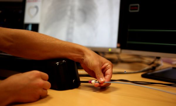
This visualization is done for simulating endovascular navigation.
 Catheter device
Insertion of a catheter in the hardware used to track the catheter motion.
 Cryoablation simulation
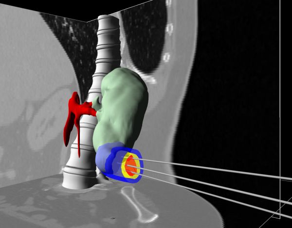
Based on GPU computing, our algorithm allows to compute the effect of cryoablation in the living tissues.
 The MIMESIS team
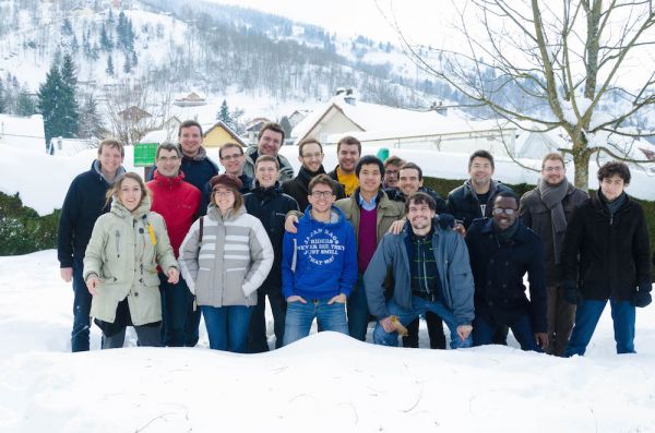
The entire MIMESIS team at the team retreat 2015 in La Bresse (Vosges, FRANCE)>.
 Cryoablation results
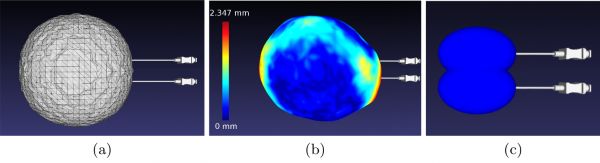
Iso-surface obtained from: (a) simulation, (b) patient-specific data (with Hausdorff dis-
tance) and (c) manufacturer.
tance) and (c) manufacturer.
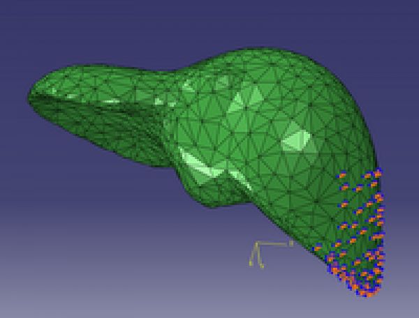 Mesh of a human liver
Liver with its boundary conditions
 Patient-specific heart geometry
Patient-specific heart geometry obtained from Cine-MRI images.
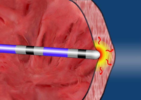 Radio-frequency ablation
For different reasons, the myocardial tissue can produce a disorder in the electrical conduction of the heart, thus causing a cardiac arrhythmia. When the arrhythmia is life-threatening, cardiologists need to ablate bthe area responsible for the pathology ased on radio-frequency (RF).
 Depolarization phase
This is the result of a simulation coupling the electrophysiology-mechanical simulation.
 Cardiac electrophysiology simulation
This image shows the depolarization times of a patient-specific heart.
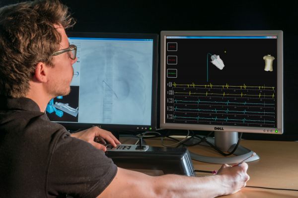 Simulator for electrocardiology training
This simulator has been developed at the end of my Ph.D. It includes two main steps: a catheter navigation in the cardiovascular system, and a second step of electrophysiology mapping. Using an hybrid (CPU-GPU) multihreaded architecture, this training system ensures a high level of interactivity and realism.
 Good times at INRIA
Once upon a time ...